
Ebola virus disease is associated with viruses of the Ebolavirus group and is characterized by severe systemic illness and a historically high case-fatality burden. Structural and sequence-based studies of Ebola virus proteins have therefore received sustained attention because viral proteins mediate entry, replication, assembly, immune evasion, and interactions with host factors. Cong, Pei, and Grishin performed predictive and comparative analyses of Ebolavirus proteins, integrating sequence variation with available three-dimensional structures and identifying putative functional regions in incompletely characterized viral proteins [1]. Such structural information provides a basis for interpreting sequence differences in relation to viral mechanisms and for identifying regions that may warrant further biochemical investigation.
The clinical importance of Ebola virus protein research is also evident from the development of therapeutic interventions. The randomized controlled trial reported by Mulangu et al. demonstrated that specific antibody-based treatments can substantially improve outcomes in Ebola virus disease [2]. Although that clinical study does not establish protein structural motifs directly, it illustrates why rigorous characterization of viral proteins and their functional regions remains relevant to therapeutic research.
Protein secondary-structure prediction has a long methodological history. Chou and Fasman developed early statistical rules for estimating secondary-structure propensity directly from amino acid sequences [3], while statistical analysis of protein-sequence patterns has also been used to characterize biologically meaningful residue distributions [4]. Earlier conformational-parameter work by Chou and Fasman quantified amino acid preferences for alpha-helical, beta-sheet, and random-coil regions [5]. These methods established the principle that local sequence composition contains information related to protein conformation.
The GOR approach formulated secondary-structure prediction using information-theoretic reasoning and local sequence context [6]. Rost, Sander, and Schneider subsequently developed the PHD system, which integrated sequence profiles and neural-network prediction [7]. Position-specific scoring matrices further improved secondary-structure prediction by incorporating evolutionary information, as demonstrated by Jones [8]; PSI-BLAST provided an efficient framework for generating such sequence profiles from iterative database searches [9].
Machine-learning methods extended these approaches by learning nonlinear relationships between sequence context and secondary structure [10]. Neural-network methods include the work of Holley and Karplus [11], Qian and Sejnowski [12], Kneller, Cohen, and Langridge [13], Malekpour et al. [14], and Qu et al. [15]. Hidden Markov and hidden semi-Markov approaches were developed by Asai et al. [16], Won et al. [17], and Aydin, Altunbasak, and Borodovsky [18]. Support-vector-machine methods were reported by Kim and Park [19], Ward et al. [20], Guo et al. [21], and Hua and Sun [22]. Fuzzy K-nearest-neighbor classification has also been applied to protein secondary-structure class prediction [23]. Collectively, these studies show that sequence-based structural prediction can be approached using statistical, evolutionary, probabilistic, and machine-learning frameworks.
The present work takes a different but complementary descriptive approach. Rather than training a predictive classifier, it partitions Ebola virus protein sequences into overlapping amino acid triplets and quantifies enrichment or depletion relative to an expected frequency. The resulting deviation parameter is normalized to allow comparison across triplets. The same principle is then applied to DSSP-derived secondary-structure categories to examine whether particular triplets are preferentially associated with alpha-helices, beta-sheets, or random structures.
The main objective is therefore to determine whether specific amino acid triplets are statistically over-represented in the analyzed Ebola virus protein sequences and whether these same triplets show distinctive secondary-structure preferences. The study does not attempt to infer biological function solely from sequence frequency. Functional interpretations in the Discussion are presented as hypotheses requiring independent structural, biochemical, or mutational validation.
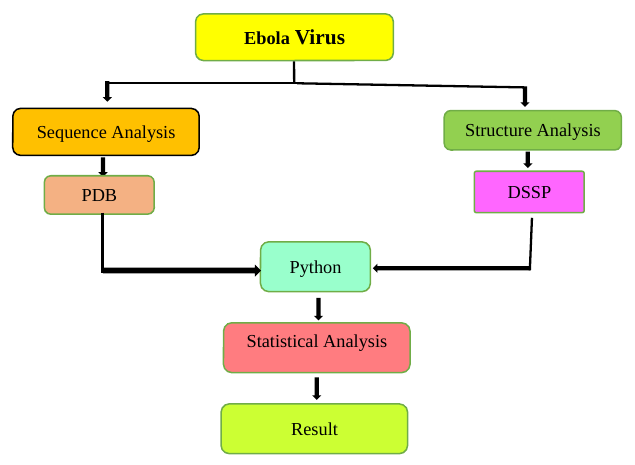
Ebola virus protein structures and associated sequences were obtained from the Protein Data Bank resource used in the study [24]. Protein sequences were processed using a Python program that enumerates overlapping amino acid triplets. For a sequence of length \(L\), the sliding triplet procedure produces \(L-2\) overlapping triplets, with consecutive windows shifted by one residue.
For each triplet, the number of occurrences in the analyzed Ebola virus sequence set was counted. The observed frequency was then calculated relative to the total number of triplets. An expected frequency was calculated using the background protein-sequence frequencies defined in the study. The observed and expected frequencies were used to derive a percentage deviation for each triplet.
This procedure provides a descriptive measure of triplet enrichment. A positive deviation indicates that a triplet occurs more frequently than expected, while a negative deviation indicates under-representation. Because raw deviations can span different numerical ranges, min–max normalization was used to place the values on a common scale between 0 and 1.
Secondary-structure assignments were obtained from DSSP, the established dictionary-of-secondary-structure procedure based on hydrogen-bonding and geometric criteria [25]. The analyzed residues were grouped into the structural categories used in this study: alpha-helix, beta-sheet, and random structure. A Python program was used to count amino acid residues and triplet occurrences associated with each structural category.
Observed and expected frequencies were calculated for the structural analysis using the same general statistical procedure as in the sequence analysis. Deviations were computed and normalized, allowing the structural preferences of selected triplets to be compared across alpha-helical, beta-sheet, and random regions.
Figure 1 summarizes the complete workflow, including sequence acquisition, triplet counting, expected-frequency calculation, deviation analysis, normalization, DSSP-based structural classification, and interpretation.
For the sequence analysis, the observed triplet frequency is defined as
where \(n_{\mathrm{Ebola}}(t)\) is the number of occurrences of triplet \(t\) in the analyzed Ebola virus protein sequences and \(N_{\mathrm{Ebola}}\) is the total number of overlapping triplets in those sequences.
The expected frequency is defined as
where \(n_{\mathrm{background}}(t)\) and \(N_{\mathrm{background}}\) refer to the corresponding triplet count and total triplet count in the background protein set used by the study.
The percentage deviation parameter is
A positive value indicates over-representation relative to the background expectation, whereas a negative value indicates under-representation. A value of zero indicates agreement between the observed and expected frequencies. The use of residue-frequency and triplet-deviation analysis is consistent with earlier statistical studies of amino acid composition and higher-order sequence patterns [26–28].
For cross-triplet comparison, the deviation parameter is normalized by min–max scaling:
Here, \(D_{\min}\) and \(D_{\max}\) are the minimum and maximum deviation values in the comparison set. The normalized value therefore lies between 0 and 1. A value near 1 identifies a triplet with a deviation close to the maximum observed in the dataset, while a value near 0 identifies a triplet close to the minimum. The same normalization procedure is applied to the secondary-structure analysis [26–28].
It is important to note that min–max normalization ranks triplets relative to the analyzed dataset; it does not by itself provide a probability, significance level, or effect-size estimate. The normalized values are therefore interpreted descriptively.
Figure 2 presents the normalized deviation values for the selected amino acid triplets identified in the Ebola virus sequence analysis.
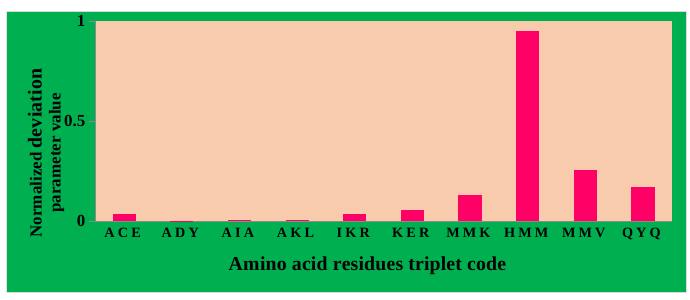
Most analyzed triplets have normalized deviation values close to the lower end of the scale, whereas a smaller subset displays markedly larger values. In the plotted results, HMM, MMV, MMM, and QYQ are among the triplets with comparatively strong deviations. These values indicate enrichment relative to the study’s background expectation; they do not, by themselves, establish conservation, essentiality, or biological function.
The biochemical composition of the enriched triplets nevertheless provides hypotheses that can be tested structurally. ACE contains alanine, cysteine, and glutamate. Cysteine can participate in disulfide bonds when positioned appropriately in an oxidizing structural environment, while glutamate can contribute to electrostatic interactions. ADY contains aspartate and tyrosine, residues that can participate in hydrogen bonding or regulatory chemistry depending on their structural context. AIA is predominantly hydrophobic and could be compatible with buried or membrane-associated environments, although sequence enrichment alone cannot determine the corresponding structural location.
AKL combines alanine, lysine, and leucine. Lysine can participate in electrostatic interactions, whereas leucine has a strong general compatibility with hydrophobic and helical environments. IKR and KER contain multiple charged residues and may therefore occur in solvent-exposed or interaction-prone regions, but the observed triplet frequency cannot identify a specific nucleic-acid-binding or localization function without independent evidence.
Methionine-rich triplets deserve careful interpretation. MMK, HMM, MMM, and MMV contain two or three methionine residues and show comparatively high deviation values in the plotted analysis. Methionine is the initiating residue in canonical protein translation, but an internal methionine-rich triplet does not imply that the corresponding region is itself a translation-initiation site. The enrichment may instead reflect local compositional characteristics of the analyzed proteins. Similarly, histidine in HMM can participate in catalytic or metal-binding environments in some proteins, but no enzymatic function can be assigned to HMM from frequency analysis alone.
QYQ combines glutamine and tyrosine. Both residues can participate in hydrogen-bonding networks, while tyrosine may undergo phosphorylation in suitable cellular contexts. These biochemical properties make QYQ structurally interesting, but experimental validation would be required before assigning a regulatory role.
Earlier triplet-frequency studies provide the statistical context for interpreting such enrichments [27], [28]. The principal result of Figure 2 is therefore the identification of non-uniform triplet usage in the analyzed Ebola virus sequences. Functional interpretations should remain secondary to that quantitative observation.
Figure 3 presents the normalized deviations of selected triplets across alpha-helix, beta-sheet, and random-structure categories.
The results indicate that several triplets exhibit relatively strong alpha-helical preferences, whereas others are associated more strongly with beta-sheet or random-structure categories. Such observations are consistent with the broader principle that amino acid propensities vary across secondary-structure environments. Position-dependent alpha-helical propensities have been demonstrated experimentally and statistically [29], and amino acid preferences for helices and beta-sheets also depend on the overall protein fold and structural context [30]. The central role of alpha-helices and beta-sheets as principal protein structural features is well established [31].
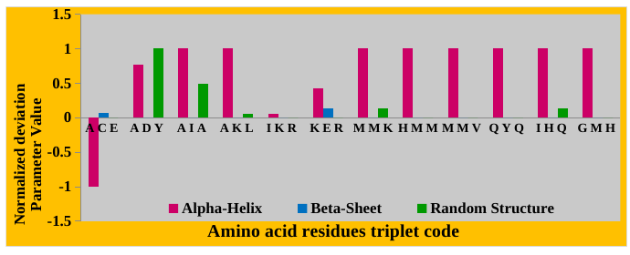
In the present analysis, AIA and AKL show relatively strong alpha-helical tendencies. This pattern is qualitatively compatible with the known helix-forming properties of alanine and leucine, although the observed triplet-level deviation should not be equated directly with an experimentally measured helix propensity. KER shows moderate representation across more than one structural class, suggesting that charged-residue context may be compatible with multiple local geometries.
MMK and other methionine-rich triplets also show pronounced alpha-helical deviations in the plotted data. HMM and MMM are among the strongest examples in the selected set. This may indicate that methionine-rich local contexts occur preferentially within helical regions of the analyzed Ebola virus structures. The result is descriptive and should be confirmed by mapping individual triplet occurrences back to specific PDB residues and protein domains.
Triplets such as ADY, IHQ, and GMH display comparatively stronger random-structure components in the analysis. Random or coil assignments can correspond to loops, linkers, turns, flexible termini, or disordered segments, but the DSSP-derived category used here does not provide a direct measure of intrinsic disorder or biological flexibility. Accordingly, the structural interpretation should remain tied to the secondary-structure classification actually analyzed.
The simultaneous use of sequence enrichment and DSSP-based structural preference is the main methodological contribution of the study. A triplet that is enriched at the sequence level and also concentrated within a specific structural category becomes a candidate for more detailed examination. Such follow-up could include residue-level mapping, solvent accessibility, evolutionary conservation, molecular dynamics, mutational analysis, or comparison across Ebolavirus species.
The normalized deviation parameter provides an intuitive ranking of triplet enrichment, but several limitations should be acknowledged. First, overlapping triplets are not statistically independent because adjacent windows share two residues. Conventional significance tests that assume independent observations would therefore require modification before being applied directly to these counts.
Second, the expected-frequency model strongly influences the deviation. If the background protein set differs substantially in amino acid composition from Ebola virus proteins, enrichment may partly reflect overall composition rather than triplet-specific organization. Future work should compare multiple background models, including independent-residue expectations, composition-matched shuffled sequences, and related viral proteins.
Third, min–max normalization depends on the extreme values in the dataset. Adding or removing a strongly enriched triplet can change the normalized values of all other triplets even if their raw frequencies remain unchanged. Reporting both raw deviation and normalized deviation would therefore improve reproducibility.
Fourth, the present structural analysis groups DSSP assignments into three broad categories. A more detailed analysis could retain individual DSSP states and distinguish alpha-helices, \(3_{10}\) helices, beta-strands, turns, bends, and unstructured states. This would provide a finer structural interpretation.
Finally, enrichment does not establish function. Claims concerning membrane association, phosphorylation, catalysis, nucleic-acid binding, viral replication, or therapeutic targeting require direct supporting evidence. The current method is most appropriately viewed as a screening strategy that identifies sequence and structure patterns for subsequent validation.
This study applies an overlapping amino acid triplet analysis to Ebola virus protein sequences and combines the resulting frequency deviations with DSSP-based secondary-structure assignments. The approach identifies non-uniform triplet usage and reveals that several selected triplets, particularly methionine-rich combinations, show comparatively strong enrichment and alpha-helical preference in the analyzed dataset.
The normalized deviation parameter provides a simple standardized measure for comparing triplets whose raw deviations differ in scale. When applied separately to sequence occurrence and secondary-structure categories, it allows candidate triplets to be prioritized for more detailed structural analysis. The results are therefore useful as a descriptive screening layer rather than as direct proof of protein function.
Future work should map enriched triplets to individual Ebola virus proteins and PDB structures, compare results across Ebolavirus species, use composition-matched sequence controls, and evaluate statistical uncertainty through permutation or resampling methods. Integrating evolutionary conservation, solvent accessibility, structural-domain annotation, and experimentally characterized functional sites would further clarify whether the identified triplet patterns have biological significance.
T.K.P. and S.A.M. contributed equally to the conceptualization, methodology, software implementation, sequence analysis, structural analysis, interpretation, visualization, manuscript preparation, and review and editing. Both authors have read and approved the final manuscript.
No specific external funding information was provided for this study.
The structural and sequence information analyzed in this study was obtained from publicly accessible PDB and DSSP resources cited in the manuscript. The derived triplet counts, processed data, and analysis scripts are available from the corresponding author upon reasonable request.
Ethical approval and informed consent were not required because this study used publicly available protein-sequence and structural data and did not involve human participants, identifiable personal data, or animal experimentation.
The authors declare no conflicts of interest.