
Cardiolipin, also referred to as cardiolipid, is a specialized phospholipid abundantly present in the cell membrane. A distinctive characteristic of cardiolipin is its occurrence primarily within the mitochondria, the powerhouse of the cells [1], [2]. The constitution of CL consists of approximately 20 percent lipids. The study of CL is essential because its structural and chemical properties [3]–[5]. CL is a dimerized lipid composed of four fatty acid chains linked to a glycerol backbone with two phosphate groups having glycerol at its third position [2], [6], [7]. This unique composition makes cardiolipin to form double layered structure within the inner mitochondrial membrane [8], [9]. The presence of cardiolipin plays a crucial role in preserving the structural integrity and ensuring the optimal functioning of mitochondria [10], [11]. It plays a crucial role in facilitating the assembly of the electron transport chain and provides stability to the respiratory complexes involved in oxidative phosphorylation [12].
Cardiolipin, a vital molecule, actively participates in cellular signalling by effectively interacting with a wide range of proteins and enzymes [13]. Cardiolipin regulates mitochondrial fusion and fission through protein binding [14]. Cardiolipin is a vital cofactor for numerous enzymes responsible for lipid metabolism and signalling pathways. Dysfunction of cardiolipin can result in major disturbances in the electron transport chain, which plays a crucial role in the effective generation of energy in the mitochondria. This disturbance ultimately results in reduced energy production and a rise in oxidative stress. The consequences of these effects can manifest as cardiac dysfunction, impacting the overall health and functioning of the heart [1]. In addition to its function as a structural component cardiolipin also plays a crucial role in various biochemical processes occurring within mitochondria [11]. When the metabolism of Cardiolipin is disrupted, it leads to harm the functional process of neurons which in turn contributes to neuronal injury [15], [16]. Cardiolipin dysfunction leads to neurological illnesses such as Alzheimer’s disease and Parkinson’s disease [13], [17].
To properly understand the mechanism of action and annotation of CL binding proteins, we must first identify the amino acid residues that interact with CL. For proteins that are still unknown, homology and sequence similarity-based approaches have shown to be ineffective. We created an CL binder technique that predicts CL binding proteins and interacting residues based solely on protein sequence, without requiring structural information.
The protein sequences needed for this study were obtained from the Protein database [18]–[20]. The selected sequences were found to be non-homologous and their structures were accurately determined at a high resolution. Our analysis focused on investigating the interactions between cardiolipin residues both interacting and non-interacting in order to gain a deeper understanding of lipid interactions [21], [22]. Residues are considered to be interacting if their atoms fall within a range of 3Å. The contact between residues and CL was accurately calculated using a Python program [23]. In this study we had taken protein sequences with similarities between 30% and 90% and their differences were taken into an account. The data set with similar protein sequences was removed and the remaining sequences were retained.
After selecting the protein, a Python programme is written with a range less than 13 Å to find the bond length [24], [25]. It will display the outcomes of amino acids rapidly reacting with the Phospholipid CL. After separating the amino acids from the results, the data was statistically evaluated [26]. The probability of amino acid distribution was found out with the help of frequency of occurrence. The frequency of occurrence of amino acid for 458 proteins of both singlet and triplet groups was calculated using the formula (1),
We used the values of the frequency of occurrence of singlet and triplet as expected count and observed count in this technique. The Deviation parameter for AA singlet and triplet in a particular structural element is computed using the formula (2),
This formula is used to determine the residue that preferentially binds to the CL as well as the amino acid that is in the “non-preferential” zone. A few proteins were chosen at random, and the procedure was carried out in the same way for all other proteins. The visualization shows the shortest distances between the amino acids that are interacting with the Phospholipid CL.
The output of the program was extracted and presented graphically.
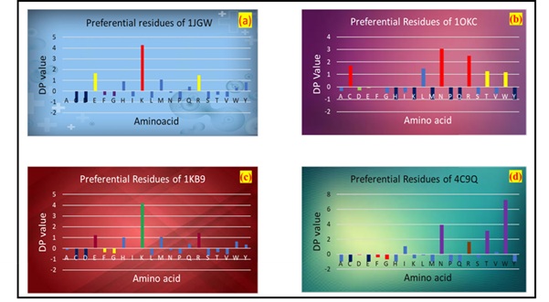
The above findings were evaluated using the Deviation parameter, which aids in determining which amino acids are favoured to bind with the ligand. We classified the distribution of amino acid into four groups namely Highly preferential, Low preferential, tends to prefer and out of range. For that we categorize the deviation parameter values into positive side and negative side. DP values in the +0.5 range are assumed to be in the preferential zone, whereas +1 or greater than 1 designates the extremely favourable sector. Similarly, DP values in the -0.5 range are considered low favoured zones, while values in the -1 range are considered out of range.
As shown in Fig for organisms like Cereibacter sphaeroides and Mus Musculus the residues such as K is the highly preferred one followed by W,H,N. Residues like E and R tends to be in the preferred zone where as residues phenylalanine and glycine have minimal probability to interact with CL and residues Cysteine and Aspartic acid are in the non-CL interacting site. For Bos Taurus residue N is highly preferred followed by R and C for organism Methanosarcina acetivorans W and N are highly preferred. Residue K being positively charged easily binds with the ligand CL. The presence of polar aliphatic positively charged amino acids was more prevalent in the CL-binding sites.
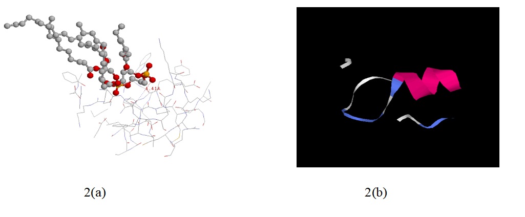
The figure above illustrates the shortest distance between the Hetatm CL and the amino acid. In this representation, the ligand is depicted in ball and stick form, while the other residues are shown in stick form and the nearest distance of the ligand and residue is around 4.41 Å. The next picture displays the structure of CL binded proteins around 13 Å.
In order to understand how CL works, we first need to know which amino acid residues bind to CL. We have created a Python program to find the binding residue. We chose Python because it is widely used and has a wide range of uses in biosciences. This method is easy and time-consuming, as it only involves a protein sequence. It allows us to perform and analyse multiple sequences at the same time, without any prior knowledge of structural data. The main finding is that the residues in the binding site of CL are highly conserved, and CL favours mixed combination of alpha helix and beta turn structure and the residues like K,W,H,N has larger propensity to bind with CL. The structural analysis shows that the nearest distance bounded by Cardiolipin is around 4.41 Å.
This study was conducted with no funding.
The authors declare that they have no conflict of interest.
The data that support the findings of this study are available in Protein Data Bank at https://doi.org/10.2210/pdbXXXX/pdb