Proteins are polypeptide structures made of one or more lengthy chains of amino acid residues [1]. The three-dimensional shape of a protein is a function of the structural organization of its atoms and the orientations of bonds within the structure. The shape of a protein plays an important role in protein properties and has been characterized in human disease context. Many techniques have investigated conformational changes under various conditions [2]. Influenza virus is an enveloped virus and is relatively susceptible to detrimental environmental conditions. In order to characterize the backbone [3] structure of amino acid residues, the phi, psi, and omega dihedral angles are employed in proteins and are characterized by every amino acid residue [4]. Because it determines its overall structure, these angles play a key role in protein structure prediction. The omega angle is defined by four synonymous backbone atoms, \(C_{i-1}, N_i, C_{\alpha i}, C_i, N_{i+1}, C_{\alpha i+1}\) [5], and the \(\omega\) angle is typically kept at \(180^\circ\). Each amino acid residue possesses a sidechain that branches off from its \(C_\alpha\) atom. Secondary structure prediction techniques have also been applied in drug design to find the conformations of small molecules [6], and to design new structures and interactions. These are basic determinations of the interatomic interactions that form the basis of molecular biology [7]. The structure and conformation of the influenza virus [8] were investigated in this study using the omega angle of amino acids. These results could be useful to biologists researching drug discovery protocols [9].
I have downloaded all the influenza virus databases from the Protein Data Bank (PDB) [10] and analyzed the torsional angles using DSSP. For our purpose, we have considered only the omega angle and utilized our own Python programming. We have chosen a selection of 127,308 non-homologous proteins from PDB, and the protein’s atomic coordination was analyzed using our own Python programming. By focusing on the omega angle, we reached a completely different conclusion that has clarified the path for future research.
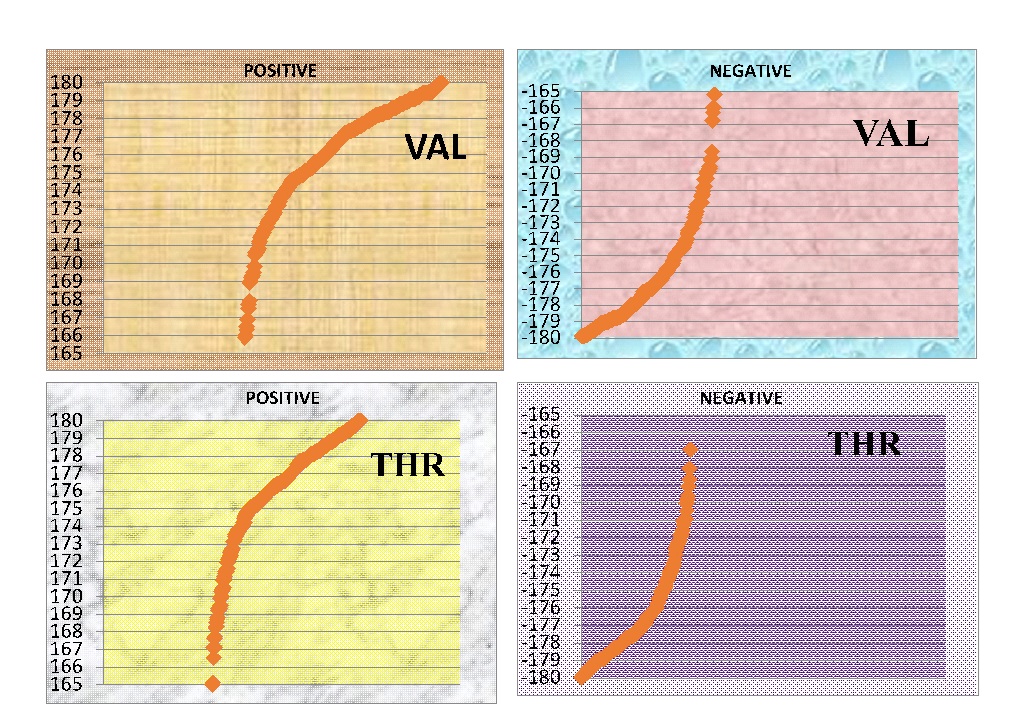
The omega angle of arginine is a particular conformation on its side chain that allows for several interactions, which are essential in determining areas of preference for this amino acid. A positive omega angle signifies a constricted conformation, while a negative omega angle is related to the positioning of arginine in protein-protein interactions, affecting amino acid preferences. There is a very small clockwise twist in the backbone in the positive omega structure of valine, which places the following amino acid at a positive rotation of the peptide bond. This is critical to protein function and has major implications for our research. In Figure 1, omega angles of glutamic acid give clues about its interactions with the conformations of neighboring residues. Positive omega angles could imply that the side chain backbone determines the acid’s behavior, while negative omega angles point to the amino acid group and are influenced by hydrogen bond orientation.
To interactions with proximate amino acids or other molecules, the conformation of a side chain to the protein backbone may be affected by omega angles. Leucine, with its branched side chain, also has its orientation altered by omega angles. This is crucial for protein structure interactions, e.g., binding sites or hydrophobic cores. Threonine, whose side chain is polar with a hydroxyl group attached, is also influenced by omega angles. In Figure 2, the orientation of the side chain can affect hydrogen bonding and other interactions critical to prot structure and functio of proteins.
This research provides crucial and extensive data regarding the influenza virus, focusing particularly on the micro-level conformational patterns of omega angles and their intricate interactions with various amino acids. The study delves into both positive and negative omega angles, offering a comprehensive description of the conformational behaviors observed in key amino acids, including ARG, GLU, LEU, VAL, and THR. By employing advanced Python programming techniques, the research meticulously analyzes these conformational variations, yielding insights that not only broaden our fundamental understanding of omega angles in the context of the influenza virus but also enhance predictive modeling capabilities. The findings from this detailed examination have significant implications for molecular biology, as they provide biologists—especially those specializing in molecular modeling—with valuable information that can refine simulation parameters, improve structural predictions, and potentially inform the development of novel therapeutic strategies. Ultimately, this work stands to contribute meaningfully to the fields of structural biology and virology by elucidating the subtle yet critical nuances of protein conformational dynamics.